Boston Children's Hospital & Neurodegenerative Imaging
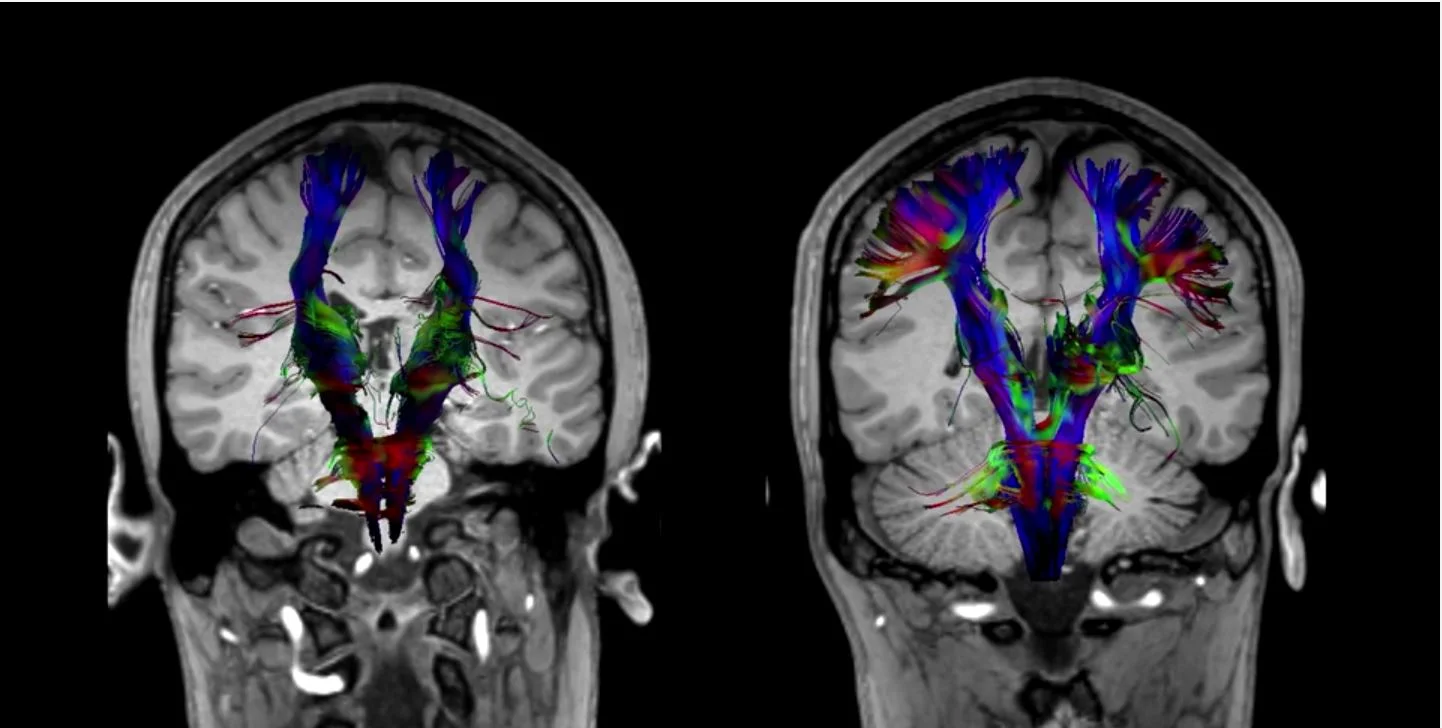
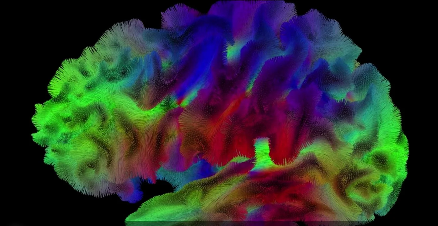
Significant advances in medical diagnostic imaging, software modernization and machine learning enables precise visualization of neural circuits, facilitating earlier diagnosis with higher confidence of neurodegenerative disorders including concussion, autism, multiple sclerosis and schizophrenia.

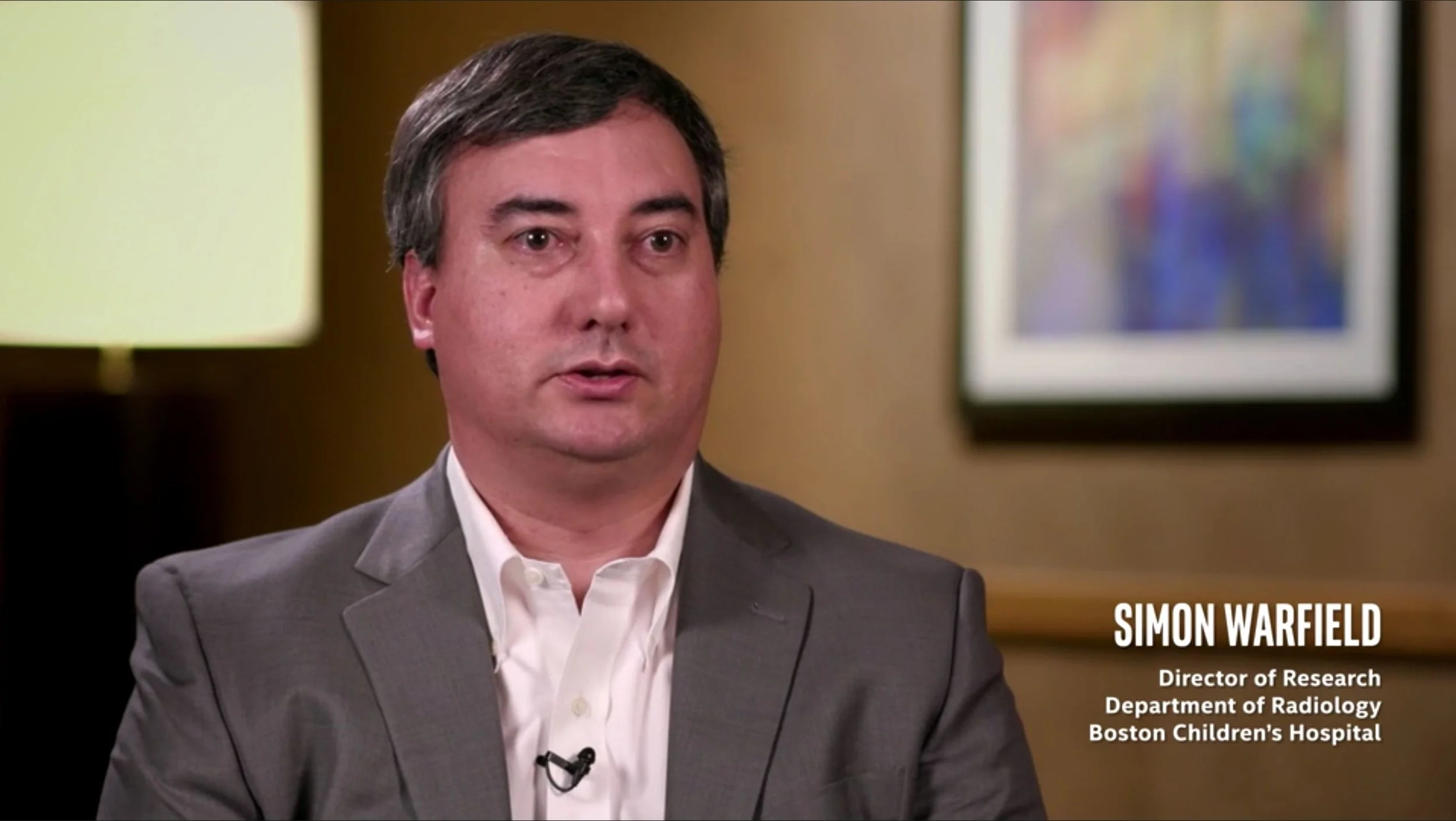
Watch this interview with Simon Warfield, N. Thorne Griscom Endowed Chair and Professor of Radiology, Harvard Medical School, Director of Radiology Research, Boston Children's Hospital
It is now recognized that a number of brain disorders, including concussion, autism and schizophrenia, are disorders of neural circuitry. However, the routine approach to diagnosis and prognosis in neurology has focused on assessing patient symptoms instead of assessing neural circuits themselves using Diffusion Compartment Imaging.

Congratulations to Boston Children’s Hospital and Harvard Medical School: Benoit Scherrer, Jaume Coll-Font, and Simon Warfield who recently won the MUSHAC competition for diffusion MRI harmonization at MICCAI 2018.
Background: Recent advances in diffusion weighted (DW) Magnetic Resonance Imaging (MRI) hardware have led to dramatic improvements of the data quality and reductions in scan duration. Such improvements come at the expense of high costs, and most clinical sites will not have access to such state-of-the-art systems in the near future.
Aims: The aim of the challenge is twofold: 1) algorithms will be tested for their ability of harmonizing the same DW MRI protocol across different MRI scanners; and 2) algorithms will be tested for their ability of enhancing the quality of routine DW MRI data sets to resemble the latest state-of-the-art acquisitions, via extrapolation of information that was not acquired given a set of training examples.
Relevance: The results of this challenge will be highly informative and clinically relevant, since the data quality from clinical scanners could potentially be improved to the same level provided by the state-of-the-art Connectome Scanner. Moreover, results will also be informative for multi-centre studies such as clinical trials, since the tested algorithms may help improve significantly their statistical power and their ability to detect differences among subject groups.
Learn about the winning algorithm:
Citation: Magn Reson Med. 2016 Sep;76(3):963-77. doi: 10.1002/mrm.25912. Epub 2015 Sep 12. Characterizing brain tissue by assessment of the distribution of anisotropic microstructural environments in diffusion-compartment imaging (DIAMOND). Scherrer B, Schwartzman A, Taquet M, Sahin M, Prabhu SP, Warfield SK.
Read about CDMRI’18: MICCAI 2018 Workshop on Computational Diffusion MRI, the MUSHAC competition and the training data provided by Cardiff University Brain Research Imaging Centre (CUBRIC).
Simon Warfield’s team recently presented the following papers at MICCAI 2018:
Citation: Stamm A., Commowick O., Menafoglio A., Warfield S.K. (2018) A Bayes Hilbert Space for Compartment Model Computing in Diffusion MRI. In: Frangi A., Schnabel J., Davatzikos C., Alberola-López C., Fichtinger G. (eds) Medical Image Computing and Computer Assisted Intervention – MICCAI 2018. MICCAI 2018. Lecture Notes in Computer Science, vol 11072. Springer, Cham. https://doi.org/10.1007/978-3-030-00931-1_9
Citation: Khan S., Rollins C.K., Ortinau C.M., Afacan O., Warfield S.K., Gholipour A. (2018) Tract-Specific Group Analysis in Fetal Cohorts Using in utero Diffusion Tensor Imaging. In: Frangi A., Schnabel J., Davatzikos C., Alberola-López C., Fichtinger G. (eds) Medical Image Computing and Computer Assisted Intervention – MICCAI 2018. MICCAI 2018. Lecture Notes in Computer Science, vol 11072. Springer, Cham. https://doi.org/10.1007/978-3-030-00931-1_4
Citation: Chatterjee S., Commowick O., Afacan O., Warfield S.K., Barillot C. (2018) Identification of Gadolinium Contrast Enhanced Regions in MS Lesions Using Brain Tissue Microstructure Information Obtained from Diffusion and T2 Relaxometry MRI. In: Frangi A., Schnabel J., Davatzikos C., Alberola-López C., Fichtinger G. (eds) Medical Image Computing and Computer Assisted Intervention – MICCAI 2018. MICCAI 2018. Lecture Notes in Computer Science, vol 11072. Springer, Cham. https://doi.org/10.1007/978-3-030-00931-1_8
Learn more about MICCAI 2018 – MEDICAL IMAGE COMPUTING & COMPUTER ASSISTED INTERVENTION
Read about the latest performance results and the implications for clinical diagnosis in "Powerful New Tools for Exploring and Healing the Human Brain"
A 10x higher image resolution (1 mm3 versus 8 mm3) increased runtimes from 16 minutes to more than 2 hours. Benchmarks showed that a four-socket Intel® Xeon® Platinum 8180 server could reduce runtimes to just 39 minutes—a 3.2X improvement versus a prior-generation, two-socket server.
Learn More
- Boston Children's Hospital, visit http://www.crl.med.harvard.edu/
- Insight Toolkit (ITK), get the source https://itk.org/ITK/resources/software.html
Acknowledgements
- Boston Children's Hospital: Simon Warfield, Benoit Scherrer, Onur Afacan, Damon Hyde, Burak Erem, Ali Gholipour
- Intel: Joseph Curley, Lisa Smith, Mike Greenfield, Alexander Bobyr, Sergey Egorov, Vadim Sherman
- Marketing & Video Production: Kathleen Ellertson, Amber Jackson, Kristine Raabe, Megan Rossman, Radhika Anand, Julie Choi
- Editorial: Rick D. Johnson, Ed Pitkin


